Serious Causes and Complications of Gallstones
Advertisement
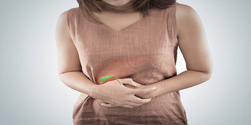
Gallstones are hardened deposits that form in the gallbladder, a small organ located beneath the liver. While they may sometimes be a symptomatic, gallstones can lead to serious medical conditions when left untreated. These include biliary colic, inflammation of the gallbladder (cholecystitis), and even infections in the bile ducts or pancreas. Understanding the causes and potential complications of gallstones is essential for early diagnosis and treatment. This guide explain the factors that contribute to gallstone formation and highlights the risks they pose to overall health.
Risk Factors and Causes
Gallstones can develop due to a combination of factors, each contributing to their formation and potential complications. Below are some of the key causes and risk factors,
Diet and Nutrition
High cholesterol diets along with insufficient fiber intake function as primary risk factors that lead to gallstone development. The high numbers of medication and empty calories from processed foods and fats trigger bile cholesterol accumulation leading to stone formation in the body. To stop gallstone development an individual must follow a diet that contains fiber along with healthy fats and fresh produce.
Obesity and Rapid Weight Loss
Gallstones become more likely in obese people because the metabolic process affecting bile salts and cholesterol becomes disrupted. The formation of gallstones may occur both from obesity and from abrupt weight reduction which leads to elevated cholesterol deposition in bile. Weight management through gradual changes in your lifestyle represents the essential approach to minimizing this risk of developing gallstones.
Genetics and Family History
Weaknesses inherited from family members increase an individual's risk for gallstones. Screening programs and precautionary steps should target individuals who carry genetic gallstone risks because these genetic factors show us how essential it is to follow monitoring and prevention methods.
Age, Gender, and Hormonal Factors
The incidence of gallstones increases in female individuals who are engage in hormone replacement therapy because estrogen levels become higher. Old age enhances the risk of gallstones because the functions of the gallbladder progressively become diminished. Efforts to prevent conditions need to account for these risk variables at the earliest stage.
Medical Conditions and Medications
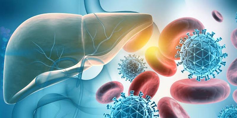
The composition of bile becomes altered when someone has diabetes or liver disease or cholesterol-lowering medications since this combination raises stone formation potential in the gallbladder. Patients who receive regular medical examinations together with proper medication adjustment can reduce their chances of developing gallstones.
Symptoms of Gallstones
Gallstones can present with a variety of symptoms, ranging from mild discomfort to severe pain, depending on their size and location.
Abdominal Pain
The main indicator of having gallstones appears as abdominal discomfort that localizes to the upper right segment of the stomach. The discomfort known as biliary colic delivers intense rapid pain which stays active between several minutes to several hours. The development of symptoms typically begins after consuming high-fat foods which causes the gallbladder to empty bile while pressing against stones inside.
Nausea and Vomiting
Gallstones trigger nausea and vomiting which mostly occur when someone experiences biliary colic symptoms. Blockages of bile cause digestion problems that produce these associated symptoms. The continuous presence of indigestion and the feeling of being bloated represents additional symptoms which appear with gallstone episodes.
Jaundice
The blockage of a bile duct by gallstones creates a condition that triggers jaundice because bile flow becomes interrupted. Essential medical help needs to be sought because these symptoms represent an advanced condition that yields yellow skin changes and eyes combined with dark urine and light colored stools.
Complications of Gallstones
Gallstones can lead to a variety of serious health complications if left untreated. Below are some of the common complications and their implications:
Gallbladder Infection (Cholecystitis)
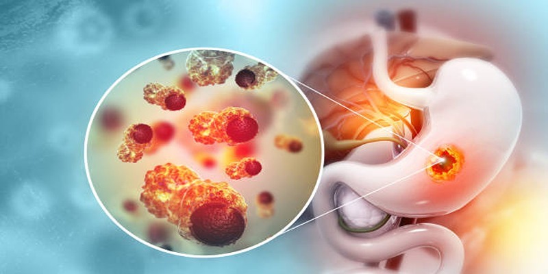
A gallstone blocking the cystic duct can cause inflammation and infection of the gallbladder, a condition known as cholecystitis. This can result in symptoms such as severe abdominal pain, fever, nausea, and vomiting. If untreated, it may lead to rupture of the gallbladder, posing a life-threatening emergency requiring surgical intervention.
Bile Duct Obstruction
When a gallstone becomes lodged in the bile duct, it can block the flow of bile from the liver to the small intestine. This obstruction often leads to jaundice, dark urine, pale stools, and intense abdominal pain. If the blockage persists, it may evolve into a serious infection of the bile duct called cholangitis, requiring immediate medical attention.
Pancreatitis
Gallstones can also enter the pancreatic duct, causing inflammation of the pancreas, a condition known as gallstone pancreatitis. This condition manifests with severe upper abdominal pain that radiates to the back, fever, rapid heartbeat, and nausea. Pancreatitis can range from mild to life-threatening and often requires hospitalization for treatment.
Gallstone Ileus
Rarely, a gallstone can enter the intestinal tract and cause a blockage, a condition known as gallstone ileus. Symptoms include severe abdominal pain, vomiting, and an inability to pass stool or gas. This condition often requires surgical intervention to remove the obstructing stone and repair the affected bowel segment.
Diagnosis and Detection
Gallstone-related complications can be effectively diagnosed and managed with a combination of clinical evaluation and diagnostic tools. Below are key methods used for diagnosis and detection:
Medical History and Physical Examination
The diagnostic process often begins with a thorough medical history and physical examination. Physicians typically inquire about the patient's symptoms, including the nature, duration, and severity of abdominal pain, nausea, or vomiting. During the physical examination, tenderness in the right upper abdomen or signs of jaundice may provide clues pointing to gallstone-related conditions.
Imaging Studies
Imaging plays a crucial role in confirming the presence of gallstones and their complications. Common imaging techniques include abdominal ultrasound, which is the first-line diagnostic tool due to its non-invasive nature and high sensitivity in detecting gallstones. Additional imaging modalities, such as CT scans or MRCP (Magnetic Resonance Cholangiopancreatography), may be used to assess complications like bile duct obstruction or gallstone ileus.
Laboratory Tests
Blood tests are essential in identifying inflammation or infection associated with gallstone complications. Elevated white blood cell counts, liver enzymes, or bilirubin levels can indicate gallstone-induced pancreatitis or bile duct blockage. These tests provide critical insights to guide further diagnostic steps.
Endoscopic Procedures
For more detailed evaluation and treatment, endoscopic techniques like ERCP (Endoscopic Retrograde Cholangiopancreatography) can be utilized. ERCP allows real-time imaging of the bile ducts and enables therapeutic interventions like the removal of obstructive stones or the placement of stents to restore bile flow.
Conclusion
Gallstone disease remains a prevalent health condition requiring timely diagnosis and management. With advancements in diagnostic imaging and endoscopic techniques, the ability to identify and treat gallstone-related complications has significantly improved. A multidisciplinary approach involving radiologists, gastroenterologists, and surgeons ensures optimal patient outcomes. Early intervention not only alleviates symptoms but also prevents severe complications such as biliary sepsis or chronic pancreatitis.
On this page
Risk Factors and Causes Diet and Nutrition Obesity and Rapid Weight Loss Genetics and Family History Age, Gender, and Hormonal Factors Medical Conditions and Medications Symptoms of Gallstones Abdominal Pain Nausea and Vomiting Jaundice Complications of Gallstones Gallbladder Infection (Cholecystitis) Bile Duct Obstruction Pancreatitis Gallstone Ileus Diagnosis and Detection Medical History and Physical Examination Imaging Studies Laboratory Tests Endoscopic Procedures ConclusionAdvertisement
Related Articles

Unnoticed Heart Attacks: Symptoms You Might Miss
By - Madison Evans

Top Best Retail Store Credit Cards for Maximum Rewards
By - Elva Flynn
Serious Causes and Complications of Gallstones
By - Kristina Cappetta

The Problem with Tanning: Myths, Risks, and Skin Safety
By - Alison Perry
Recognizing Symptoms of Ehlers-Danlos Syndrome
By - Maurice Oliver
Home Appraisals Made Simple: A Must-Know Guide for Homeowners
By - Georgia Vincent

Discover Yorkshire’s Top Seaside Towns for a Relaxing Getaway
By - Tessa Rodriguez

Refinancing Your Home Loan: What You Need to Know
By - Vicky Louisa

Four Beneficiaries of Wealth and Their Roles in Society
By - Pamela Andrew

Top Strategies for Keeping Your Investment Portfolio Aligned
By - Elva Flynn
Subic 2 Days 1 Night Guide: Fun Activities and Must-Visit Places
By - Sean William

How a Concentrated Stock Position Can Affect Your Portfolio
By - Georgia Vincent